If you are interested in products and services related to the research phase in this field, please contact for further inquiries.
Magnetic Resonance Imaging (MRI) has long been a cornerstone of modern medical diagnostics, offering unparalleled insights into the human body's internal structures. However, traditional MRI systems come with significant limitations, including high costs, large footprints, and the need for specialized facilities. The advent of portable low-field MRI (LF-MRI) has the potential to revolutionize neuroimaging by addressing these challenges. This article delves into the technological advancements, clinical applications, and future prospects of portable LF-MRI, highlighting its transformative impact on the field of neuroimaging.
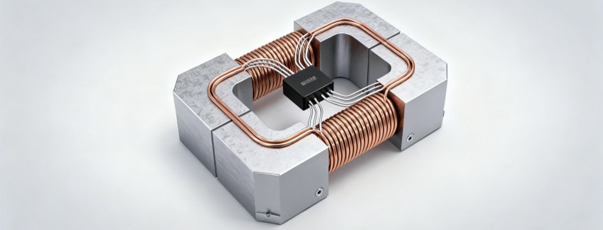
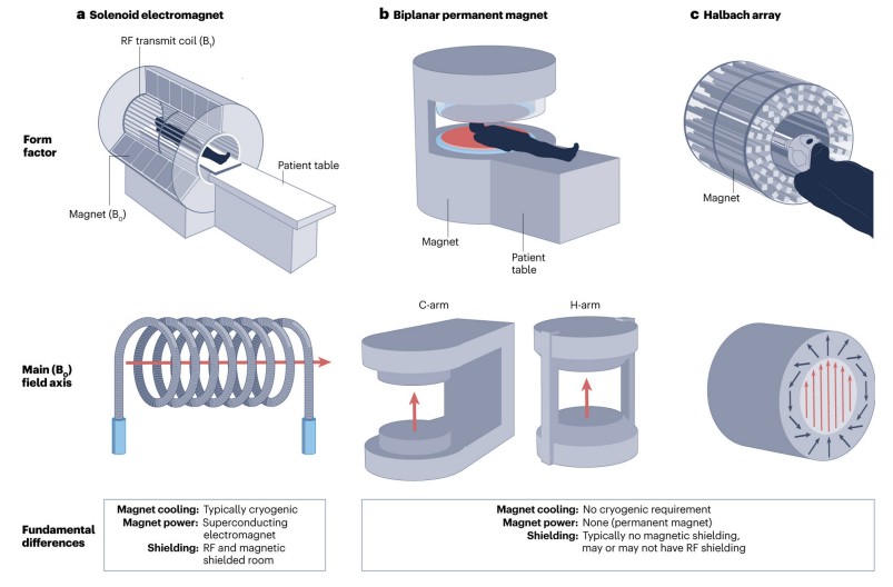 Fig.1 Types of MRI geometries for portable LF-MRI. (Kimberly W. T., et al., 2023)
The Evolution of MRI Technology
MRI technology has evolved significantly since its inception in the early 1980s. Initially, MRI machines operated at low magnetic field strengths, typically between 0.05 and 0.35 Tesla (T). These early systems laid the groundwork for more advanced imaging techniques but were limited by their lower signal-to-noise ratio (SNR), which resulted in less detailed images. In response, the industry shifted towards higher field strengths, with systems operating at 1.5T and above becoming the norm. These high-field MRI systems offered superior image resolution and diagnostic capabilities, making them the preferred choice for clinical applications.
However, the high costs, large size, and operational complexities of high-field MRI systems limited their accessibility. Many healthcare facilities, particularly in resource-constrained environments, could not afford to install and maintain these advanced systems. This limitation highlighted the need for more portable and affordable imaging solutions. The resurgence of interest in low-field MRI (LF-MRI) has been driven by advancements in technology that have mitigated the earlier limitations of these systems. The first FDA-approved portable LF-MRI device was deployed in 2020, marking a significant milestone in the evolution of MRI technology. Since then, LF-MRI has been increasingly investigated for various clinical indications and environments, demonstrating its potential to democratize MRI access globally.
Technological Innovations in Portable LF-MRI
Electromagnetic Noise Cancellation
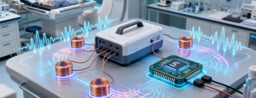
One of the key challenges in portable LF-MRI is managing electromagnetic interference (EMI). Unlike traditional MRI systems, which are housed in shielded rooms, portable LF-MRI devices often operate in unshielded environments. This exposes them to various sources of EMI, which can degrade image quality. Recent advancements in active EMI cancellation methods have addressed this challenge. Techniques such as the use of EMI sensing coils and deep learning algorithms have enabled portable LF-MRI systems to effectively cancel out EMI, ensuring high-quality imaging in diverse settings.
Machine Learning and AI
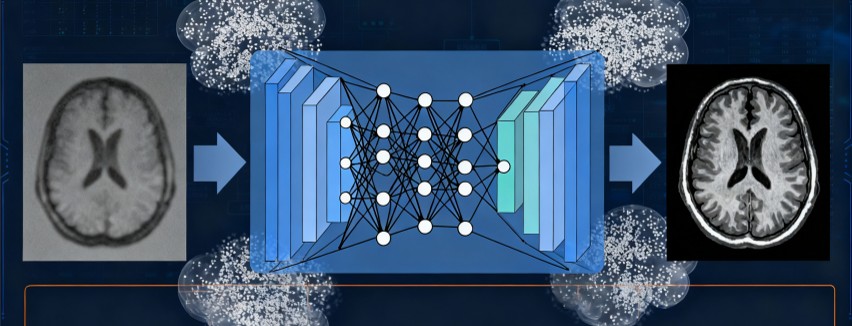
Machine learning and artificial intelligence (AI) have played a crucial role in enhancing the capabilities of LF-MRI. Convolutional neural networks (CNNs) and other AI algorithms have been employed to improve image quality and resolution. These algorithms can predict high-resolution images from low-resolution inputs, effectively bridging the gap between LF-MRI and high-field MRI. By leveraging the power of AI, LF-MRI systems can now produce diagnostic-quality images that were previously unattainable with lower field strengths.
Pulse Sequences and Data Acquisition
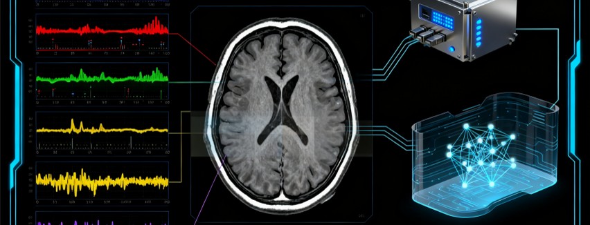
Traditional MRI protocols, such as T1-weighted (T1W), T2-weighted (T2W), fluid-attenuated inversion recovery (FLAIR), diffusion-weighted imaging (DWI), and susceptibility-weighted imaging (SWI), have been adapted for LF-MRI. New pulse sequences and data acquisition methods, such as TrueFISP-based MR fingerprinting and deep learning reconstruction, have further improved image quality. These advancements have enabled LF-MRI systems to produce detailed images of the brain and other neurological structures, making them suitable for a wide range of clinical applications.
Hardware Innovations
Advances in magnet design, gradient coil technology, and RF coil engineering have led to lighter, more efficient, and more homogeneous magnets. The use of permanent magnets, such as those made from neodymium–iron–boron (NdFeB) or samarium–cobalt (SmCo), has eliminated the need for supercooling cryogens and reduced power consumption. These magnets are typically arranged in C-arm, H-arm, or Halbach array geometries, which allow for more compact and portable designs. Additionally, the use of materials like Litz wire and ultra-low-noise RF preamps has contributed to improved detector performance, further enhancing the capabilities of LF-MRI systems.
Clinical Applications of Portable LF-MRI
- Stroke and Intracerebral Haemorrhage
Timely diagnosis is crucial in managing stroke and intracerebral haemorrhage (ICH). Portable LF-MRI systems have demonstrated high sensitivity and specificity in detecting acute ischaemic stroke (AIS) and ICH, comparable to conventional high-field MRI. These systems can be deployed in emergency departments, ambulances, and remote areas, facilitating rapid diagnosis and treatment. For example, a study published in Science Advances demonstrated that LF-MRI could detect diffusion restriction in 90% of patients with acute ischaemic stroke identified on high-field MRI. This capability makes LF-MRI an invaluable tool in emergency settings, where rapid diagnosis can significantly impact patient outcomes.
- Cardiac Arrest and Critical Care
Critically ill patients often require neuroimaging to monitor for complications such as hypoxic-ischaemic brain injury. Portable LF-MRI can be used at the bedside, allowing for continuous monitoring without the need to transport patients to centralized imaging suites. This has been particularly beneficial in intensive care units (ICUs) and neonatal ICUs (NICUs), where patient transport can pose significant risks. For instance, a study published in Resuscitation reported that LF-MRI could detect hypoxic-ischaemic injury in cardiac arrest survivors, providing critical information for neurological prognostication.
- Paediatric Brain Development
Neuroimaging plays a crucial role in understanding paediatric brain development and diagnosing conditions such as hydrocephalus. LF-MRI has been successfully used in paediatric populations, including neonates, without the need for sedation. This has significant implications for early diagnosis and intervention in resource-constrained settings. For example, a study published in Archives of Disease in Childhood demonstrated that LF-MRI could produce high-quality images of the neonatal brain, facilitating the diagnosis of hydrocephalus and other neurological conditions.
- Chronic Neurological Diseases
LF-MRI has potential applications in monitoring chronic neurological conditions such as multiple sclerosis (MS) and Alzheimer's disease. The ability to perform serial imaging at the point of care could improve disease management and treatment monitoring. For instance, a study published in NeuroImage: Clinical reported that LF-MRI could sensitively detect and accurately quantify MS lesions, making it a valuable tool for monitoring disease progression.
Future Prospects and Challenges
Image Quality
The inherently lower resolution and SNR of LF-MRI require further advancements in image enhancement and reconstruction techniques to match the diagnostic capabilities of high-field MRI. Ongoing research is focused on developing new algorithms and hardware innovations to improve image quality and reduce acquisition times.
Clinical Validation
Formal validation studies are needed to establish the clinical utility of LF-MRI across a range of environments and patient populations. This includes assessing the effectiveness of LF-MRI in detecting subtle pathologies and its impact on patient outcomes. Collaborative efforts between researchers, clinicians, and industry partners are essential to gather robust clinical data and ensure the widespread adoption of LF-MRI.
Safety and Regulatory Considerations
Ensuring the safety and regulatory compliance of LF-MRI in diverse settings is crucial. This includes addressing potential biases in AI algorithms and ensuring data privacy and security. Regulatory bodies such as the FDA and EMA must establish clear guidelines for the use of LF-MRI in clinical practice, ensuring that these systems meet the highest standards of safety and efficacy.
Global Accessibility
Deploying LF-MRI in resource-constrained environments requires addressing logistical challenges such as equipment storage, operational logistics, and infrastructure stability. Local community engagement and training are essential for successful implementation. By working closely with local healthcare providers and communities, LF-MRI can be effectively deployed to improve access to neuroimaging in underserved regions.
Conclusion
Portable low-field MRI (LF-MRI) represents a significant advancement in neuroimaging, offering a more accessible and affordable alternative to conventional high-field MRI. With ongoing technological innovations and clinical validation, LF-MRI has the potential to transform the diagnosis and management of neurological conditions worldwide. As this technology continues to evolve, it is poised to democratize MRI access, making high-quality neuroimaging available to a broader population. The future of LF-MRI looks promising, with the potential to revolutionize neuroimaging and improve patient outcomes across diverse clinical settings.
If you have related needs, please feel free to contact us for more information or product support.
Reference
- Kimberly, W. Taylor, et al. "Brain imaging with portable low-field MRI." Nature reviews bioengineering 1.9 (2023): 617-630.
This article is for research use only. Do not use in any diagnostic or therapeutic application.