- Home
- Resource
- Disease Diagnosis
- Cardiovascular Diseases
- The Stroke Diagnostic Toolbox: From FAST Assessment to Advanced Biomarkers
- Home
- IVD
- By Technology Types
- By Diseases Types
- By Product Types
- Research
- Resource
- Distributors
- Company

Stroke is a time-critical medical emergency caused by interrupted blood flow to the brain, requiring rapid diagnosis to prevent irreversible damage and improve patient outcomes. This resource page provides a comprehensive overview of the modern stroke diagnostic pathway, detailing the integrated use of clinical assessments like the FAST test, advanced imaging technologies such as CT and MRI, essential laboratory testing, and the promising role of emerging blood-based biomarkers to guide effective and timely clinical decision-making.
A stroke is a life-threatening medical emergency that occurs when the blood supply to part of the brain is interrupted or severely reduced, depriving brain tissue of oxygen and nutrients. Within minutes, brain cells begin to die. There are two primary types: ischemic stroke, caused by a blockage in an artery, and hemorrhagic stroke, caused by a bleeding blood vessel in or around the brain. The consequences are often rapid and devastating, potentially leading to permanent brain damage, long-term disability, or death. Recognizing the sudden signs—such as face drooping, arm weakness, and speech difficulty—and seeking immediate medical attention is critical, as prompt diagnosis and treatment are paramount to saving brain function and improving recovery outcomes.
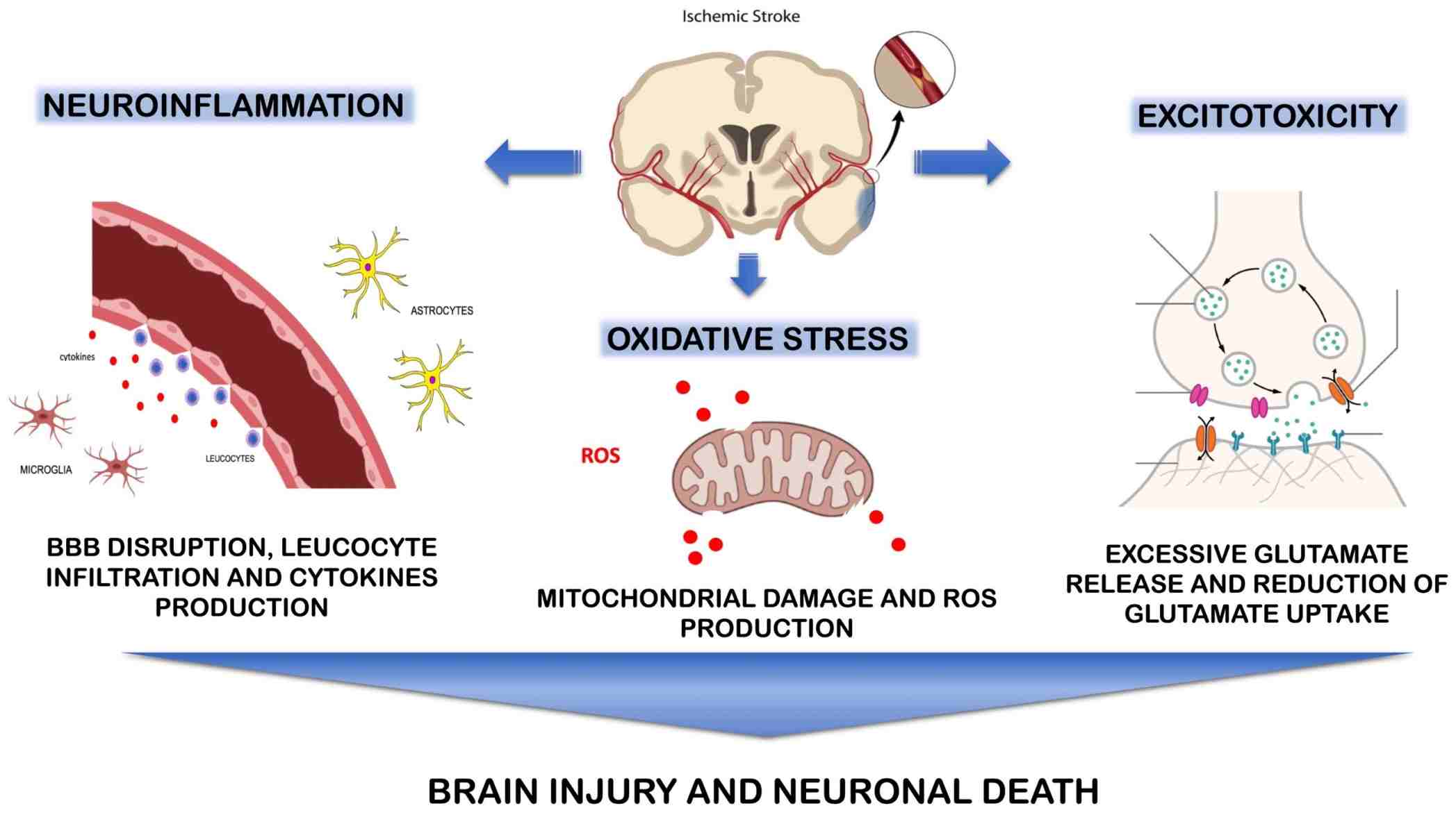 Fig.1 A summary of the pathophysiology involved in ischemic stroke. (Maida C D, et al., 2024)
The FAST assessment is a simple, widely promoted tool designed for the rapid identification of a suspected stroke by the public and first responders. It is a mnemonic that stands for Face drooping, Arm weakness, Speech difficulty, and Time to call emergency services. It works by checking for unilateral facial numbness or an uneven smile, arm weakness or drift when both arms are raised, and slurred or strange speech. The critical component is "Time," emphasizing that any one of these signs warrants an immediate emergency response, as every minute of delay in treatment leads to greater brain damage. While not a diagnostic tool for healthcare professionals, its purpose is to bridge the gap between symptom onset and the hospital, triggering the rapid activation of the emergency stroke system.
Brain imaging is the critical first step in diagnosing a stroke, serving to rapidly confirm the event, distinguish between an ischemic stroke (caused by a clot) and a hemorrhagic stroke (caused by bleeding), and guide immediate treatment decisions. The primary goal is to quickly obtain actionable information to preserve brain function, leading to a standardized protocol that often begins with the following key modalities.
Non-Contrast Computed Tomography (CT) Scan
The non-contrast CT scan is the initial and most rapidly acquired imaging test in the emergency setting. Its primary and most crucial role is to rule out intracranial hemorrhage, as this finding would contraindicate clot-busting drugs (thrombolytics). While it can appear normal in the very early stages of an ischemic stroke, it can reveal subtle early signs like a hyperdense artery sign or loss of gray-white matter differentiation.
Magnetic Resonance Imaging (MRI)
MRI, particularly the diffusion-weighted imaging (DWI) sequence, is the gold standard for detecting acute ischemic stroke. DWI can identify irreversibly injured brain tissue within minutes of symptom onset, making it highly sensitive for diagnosing ischemic strokes that may be invisible on a CT scan. It is also superior for identifying stroke mimics and can be used to detect hemorrhage with specific sequences.

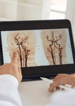
Vascular Imaging
Vascular imaging, including CT angiography (CTA) and MR angiography (MRA, focuses on visualizing the blood vessels in the neck and brain. Its key purpose is to identify the location and extent of a blockage in cases of ischemic stroke, such as a large vessel occlusion, which is essential for determining eligibility for mechanical thrombectomy. In hemorrhagic stroke, it can help locate the source of bleeding, such as an aneurysm or arteriovenous malformation.
While brain imaging confirms a stroke and defines its type, laboratory testing provides essential supportive information to guide management and uncover the underlying cause. Traditional tests are crucial for ruling out stroke mimics and assessing patient health, but the emerging field of stroke biomarkers promises to revolutionize diagnosis by offering rapid, blood-based tools to quickly confirm brain injury, differentiate stroke types, and even predict outcomes.
Traditional Laboratory Testing
Initial laboratory workup is essential to support diagnosis, rule out stroke mimics, and guide safe treatment selection.
Emerging Blood-Based Biomarkers
Emerging blood-based biomarkers offer the potential to provide rapid, specific diagnostic and prognostic information about the brain injury itself, directly from a blood sample.
To advance the precision diagnosis of stroke, Alta DiagnoTech is dedicated to transforming patient care through a comprehensive portfolio of innovative in vitro diagnostic (IVD) solutions. Our products are engineered to integrate seamlessly into the acute clinical workflow, delivering rapid and reliable results that empower healthcare professionals to make faster, more confident decisions—from initial diagnosis and differential assessment to prognostic evaluation, ultimately improving patient outcomes. If you have related needs, please feel free to contact us for more information or product support.
| Product Name | Technology | Application |
| Rapid Stroke Triage Panel (GFAP/NDKA) | Lateral Flow Immunoassay (LFIA) | Point-of-care qualitative detection of key biomarkers to aid in initial stroke assessment and triage. |
| High-Sensitivity Cardiac Troponin I Assay | Chemiluminescent Immunoassay (CLIA) | Quantitative measurement of cardiac troponin-I to identify concurrent myocardial infarction and assess cardio-embolic stroke risk. |
| GFAP Quantification Assay Kit | Electrochemiluminescence Immunoassay (ECLIA) | Quantitative measurement of Glial Fibrillary Acidic Protein (GFAP) in plasma/serum to aid in the differentiation of intracerebral hemorrhage from ischemic stroke. |
| Automated Coagulation Panel (PT/INR, aPTT) | Photometric & Mechanical Clot Detection | Comprehensive evaluation of coagulation status to guide thrombolytic therapy and manage anticoagulation. |
| Ischemic Stroke Biomarker Panel (NDKA/S100B) | Multiplex Immunoassay | Simultaneous quantitative measurement of a panel of biomarkers to support the diagnosis of acute ischemic stroke and provide prognostic information. |
Reference
This article is for research use only. Do not use in any diagnostic or therapeutic application.
|
There is no product in your cart. |
Copyright © 2026 Alta DiagenoTech. All rights reserved.