- Home
- Resource
- Disease Diagnosis
- Endocrine Diseases
- The Diagnostic Pathway for Paget's Disease of Bone: Integrating Biomarkers and Imaging
- Home
- IVD
- By Technology Types
- By Diseases Types
- By Product Types
- Research
- Resource
- Distributors
- Company

Paget's disease of bone is a chronic focal skeletal disorder characterized by accelerated and disorganized bone remodeling. This resource outlines a clear, integrated diagnostic pathway for the condition, detailing how to effectively combine biochemical markers of bone turnover with definitive imaging studies. It presents the systematic steps from initial suspicion through confirmation and disease assessment, concluding with an overview of the specialized in vitro diagnostic (IVD) tools that support precise detection and monitoring throughout the patient journey.
Paget's disease of bone is a chronic, focal skeletal disorder characterized by abnormally accelerated and disorganized bone remodeling, leading to enlarged, deformed, and mechanically weakened bones. This condition results from an imbalance between excessive bone resorption by overactive osteoclasts and compensatory, chaotic new bone formation by osteoblasts. While often asymptomatic and discovered incidentally, it can cause bone pain, deformities (such as bowed legs or an enlarged skull), arthritis, fractures, and neurological complications if it affects the skull or spine. Diagnosis hinges on recognizing its characteristic radiographic features and elevated biochemical markers of bone turnover.
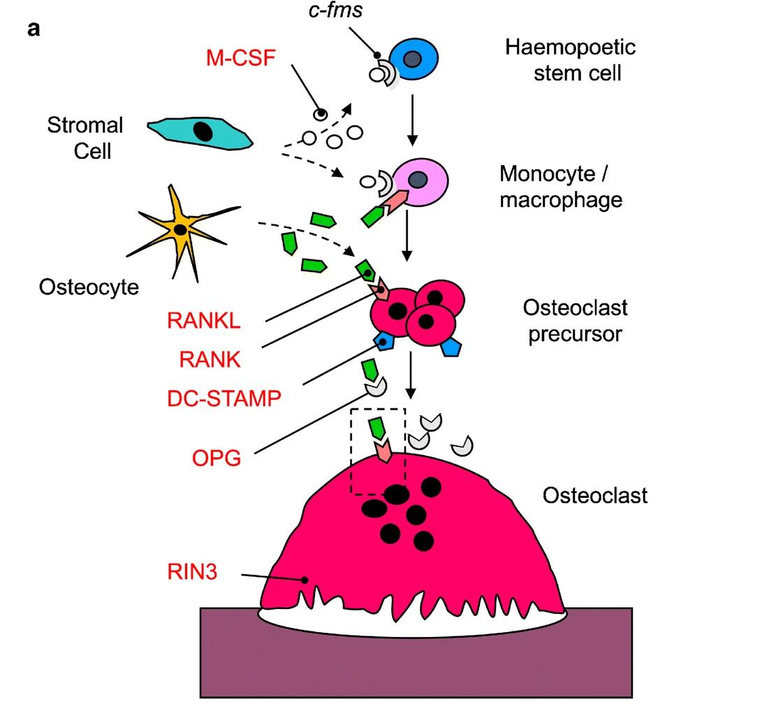 Fig.1 Genes implicated in Paget's disease and related syndromes. (Navnit S. Makaram & Stuart H. Ralston., 2021)
Biomarkers of bone turnover are crucial tools in the diagnosis and management of Paget's disease of bone, providing a non-invasive measure of the condition's accelerated and dysregulated remodeling activity. These serum and urine tests reflect the rate of bone formation and resorption, serving to raise initial suspicion, quantify disease burden, and monitor therapeutic response.

Serum Alkaline Phosphatase (ALP)
Serum total alkaline phosphatase (ALP) is the most widely used and sensitive screening biomarker for Paget's disease. It primarily reflects the elevated osteoblastic bone formation activity that follows the initial wave of excessive resorption. A persistently elevated ALP level on a routine chemistry panel is a common incidental finding that should prompt further investigation for Paget's disease, as its degree often correlates with the extent and activity of skeletal involvement.

Specificity and Confirmation
While total ALP is a sensitive screen, it lacks specificity, as levels can also be elevated in hepatobiliary disease. To confirm a skeletal source, bone-specific alkaline phosphatase (BALP) provides greater specificity. In complex cases, such as monostotic disease or for monitoring potent therapy, bone resorption markers like serum C-telopeptide (CTX) or urinary N-telopeptide (NTX) can offer additional, complementary information on the resorptive component of the turnover cycle.
Imaging studies are essential for confirming the diagnosis and assessing the extent of Paget's disease of bone. While biochemical markers indicate increased metabolic activity, imaging provides direct visualization of the characteristic structural changes. A combined approach using two primary modalities—each with a distinct role—forms the cornerstone of radiologic evaluation.

Plain radiography (X-ray) is the gold standard for definitive diagnosis. It reveals the pathognomonic structural abnormalities of Paget's disease, including bone enlargement, cortical thickening, trabecular coarsening, and the classic lytic "flame-shaped" or "blade of grass" lesions in long bones, as well as the "cotton wool" appearance in the skull. These specific findings are often diagnostic in the appropriate clinical context.

Radionuclide bone scan (bone scintigraphy) is the most sensitive tool for determining the full anatomic extent of skeletal involvement. It identifies all metabolically active sites, effectively distinguishing between monostotic (single bone) and polyostotic (multiple bones) disease. Its primary role is to serve as a "roadmap," guiding targeted X-rays to active areas for definitive morphologic confirmation.
The diagnosis of Paget's disease follows a logical, integrated pathway that systematically translates clinical clues into a confirmed diagnosis. This process can be summarized in five key steps, each building upon the last to provide a complete picture of disease activity and anatomy.
As a specialized in vitro diagnostic (IVD) provider for Paget's disease of bone, Alta DiagnoTech offers a comprehensive portfolio of product solutions. Our high-precision assays enable accurate identification of abnormal bone turnover, confirmation of disease activity, and continuous monitoring of therapeutic response. These reliable testing tools form an essential foundation for the integrated diagnostic strategy that combines biochemical analysis with imaging confirmation. If you have related needs, please feel free to contact us for more information or product support.
| Product Name | Technology | Application |
| Alkaline Phosphatase (ALP) Assay Kit | Colorimetric / Enzymatic | Quantitative measurement of total serum ALP; the primary screening test for detecting elevated bone turnover suggestive of Paget's disease. |
| Bone-Specific Alkaline Phosphatase (BALP) Immunoassay Kit | Chemiluminescent Immunoassay (CLIA) | Specific measurement of the bone-derived ALP isoenzyme to confirm skeletal origin of elevated ALP and enhance diagnostic specificity. |
| C-Telopeptide (CTX) Serum Assay Kit | Electrochemiluminescence Immunoassay (ECLIA) | Quantitative detection of type I collagen degradation products; a sensitive marker of bone resorption used for assessing high turnover states and monitoring bisphosphonate therapy response. |
| N-Telopeptide (NTX) Urine Assay Kit | Enzyme-Linked Immunosorbent Assay (ELISA) | Measurement of cross-linked N-telopeptides in urine; an alternative bone resorption marker for monitoring disease activity and treatment efficacy. |
| Liver Function Profile (GGT, ALT, AST) Assay Panel | Colorimetric / UV-Enzymatic | Differential diagnosis panel to rule out hepatobiliary disease as a cause of elevated total ALP, ensuring accurate interpretation of bone turnover markers. |
| Automated Bone Turnover Panel (ALP + CTX) | CLIA / ECLIA Integrated Platform | Simultaneous assessment of bone formation (ALP) and resorption (CTX) activity, providing a comprehensive baseline and monitoring profile for Paget's disease management. |
Reference
This article is for research use only. Do not use in any diagnostic or therapeutic application.
|
There is no product in your cart. |
Copyright © 2026 Alta DiagenoTech. All rights reserved.