Diagnosis and monitoring of multiple sclerosis (MS) has long relied on clinical assessment, MRI, and invasive cerebrospinal fluid (CSF) analysis, which face challenges in sensitivity, specificity, and accessibility. Next-generation biomarkers such as neurofilament light chain (NfL), chitinase-3-like-1 (CHI3L1), and microRNAs are revolutionizing medical care, enabling earlier detection, precise subtyping, and objective treatment tracking.
Introduction to Multiple Sclerosis (MS)
Multiple sclerosis (MS) is a chronic autoimmune disease of the central nervous system (CNS) characterized by inflammation, demyelination, and neurodegeneration. It affects over 2.8 million people worldwide, with symptoms ranging from vision problems and fatigue to mobility impairment and cognitive decline.
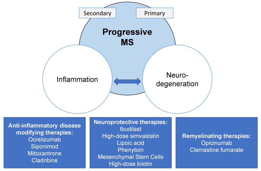
 Fig.1 Progressive multiple sclerosis (MS) is caused by the interaction of inflammation and neurodegeneration. (Macaron G, Ontaneda D., 2019)
Traditional Methods for Multiple Sclerosis (MS) Diagnostics
The diagnosis of multiple sclerosis (MS) has historically relied on a combination of clinical assessment and paraclinical tests to demonstrate dissemination in time (DIT) and dissemination in space (DIS). The following traditional methods remain cornerstone approaches in MS diagnostics:
- Magnetic Resonance Imaging (MRI)
MRI is the cornerstone of MS diagnosis, detecting characteristic CNS lesions through T2-weighted/FLAIR sequences (showing plaques) and gadolinium-enhanced T1 imaging (revealing active inflammation). While non-invasive and highly sensitive for spatial dissemination (DIS), it has limitations in specificity (mimics like NMOSD) and accessibility in resource-limited settings.
- Cerebrospinal Fluid (CSF) Examination
CSF analysis, particularly oligoclonal bands (OCBs), provides critical evidence of intrathecal IgG synthesis, supporting MS diagnosis when clinical/MRI findings are inconclusive. Though highly specific, its invasiveness (lumbar puncture) and discomfort limit routine use, driving demand for blood-based alternatives.
- Visually Evoked Potentials (VEPs) & Optical Coherence Tomography (OCT)
VEPs identify delayed visual conduction (e.g., optic neuritis), while OCT measures retinal nerve fiber layer thinning, reflecting neurodegeneration. These tools offer objective, non-invasive insights but lack disease specificity and focus primarily on anterior visual pathway damage.
Significance of Biomarkers in Multiple Sclerosis (MS) Diagnostics
Biomarkers are transforming multiple sclerosis (MS) diagnostics by overcoming key limitations of conventional methods like MRI and cerebrospinal fluid analysis. They play a pivotal role in enabling earlier and more accurate diagnosis through objective, quantifiable measures of neuroaxonal damage and neuroinflammation. Compared to traditional approaches, biomarkers offer significant advantages:
- Early Detection: Potential to identify disease before clinical symptoms appear
- Objective Monitoring: Quantitative measures of disease activity and progression
- Reduced Invasiveness: Blood-based biomarkers minimizing need for lumbar punctures
- Subtype Differentiation: Helping distinguish between MS variants (RRMS, PPMS, SPMS)
- Treatment Response Assessment: Monitoring therapeutic efficacy and safety
Emerging Biomarkers Revolutionizing Multiple Sclerosis (MS) Diagnosis
Emerging biomarkers provide objective indicators that can complement existing methods, allowing for earlier detection and personalized treatment strategies. Three particularly promising biomarkers include:
Neurofilament Light Chain (NfL)
As a sensitive marker of axonal damage, NfL has emerged as a cornerstone in MS biomarker research. Elevated levels in cerebrospinal fluid (CSF) and blood correlate strongly with disease activity, making it valuable for monitoring relapses, progression, and treatment efficacy.
Chitinase-3-like-1 (CHI3L1)
CHI3L1 levels reflect glial activation and neuroinflammation, serving as both a diagnostic aid and prognostic indicator. Its ability to predict conversion from relapsing-remitting to progressive disease makes it especially valuable for clinical decision-making and trial stratification.
MicroRNAs
Specific miRNA signatures (e.g., miR-150, miR-21) demonstrate disease-specific expression patterns that may enable earlier diagnosis and subtype differentiation. Their stability in biofluids and potential for multi-analyte panels position them as promising tools for precision medicine approaches in MS.
IVD Development Based on Multiple Sclerosis (MS) Biomarkers
With the breakthrough in the research of multiple sclerosis (MS) biomarkers, the field of in vitro diagnostics (IVD) is welcoming new product development opportunities. Using biomarkers such as NfL, CHI3L1 and microRNAs, new IVD products can be developed to promote early and accurate diagnosis of MS.
Serum/Plasma NfL Quantification Kits
Ultra-sensitive immunoassays (e.g., Simoa, ELISA) for measuring neurofilament light chain in blood enable minimally invasive monitoring of MS disease activity and treatment response, with potential for integration into automated chemiluminescence platforms.
CHI3L1 Rapid Testing Systems
These quantitative immunoassays detect CHI3L1 protein levels in serum/plasma, specifically targeting the neuroinflammatory component of progressive MS. Utilizing lateral flow or microfluidic technology, the POCT-optimized systems provide rapid results.
microRNA Multi-Analyte Panels
qPCR- based or NGS-based diagnostic panels targeting MS-specific miRNA signatures (e.g., miR-150, miR-21) may enable subtype stratification and early detection through liquid biopsy approaches. The panel has high stability in extracellular vesicles and enables multi-analyte detection.
Future of Biomarker-Based Diagnostics in Multiple Sclerosis (MS)
While biomarkers like neurofilament light chain (NfL), CHI3L1, and microRNAs have revolutionized multiple sclerosis (MS) diagnosis and monitoring, their clinical application still faces challenges in standardization, sensitivity, and specificity. Looking ahead, the integration of multi-omics technologies and artificial intelligence will drive the development of precision diagnostic systems that combine multimodal biomarkers, imaging, and clinical data, ultimately advancing early detection, personalized treatment, and prognosis assessment in MS.
Alta DiagnoTech is committed to pioneering advanced in vitro diagnostic (IVD) solutions for autoimmune disorders like multiple sclerosis (MS). Empowering early and accurate detection, our products are at the forefront of precision healthcare. If you have related needs, please feel free to contact us for more information or product support.
References
- Macaron G, Ontaneda D. Diagnosis and management of progressive multiple sclerosis[J]. Biomedicines, 2019, 7(3): 56.
- Ford H. Clinical presentation and diagnosis of multiple sclerosis[J]. Clinical Medicine, 2020, 20(4): 380-383.
This article is for research use only. Do not use in any diagnostic or therapeutic application.


 Fig.1 Progressive multiple sclerosis (MS) is caused by the interaction of inflammation and neurodegeneration. (Macaron G, Ontaneda D., 2019)


